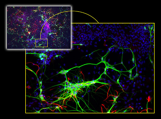
A culture dish containing many thousands of cells from the brain of a rat (top left), labelled with fluorescent dyes. The image covers a very large area – 6mm in diameter. The inset demonstrates the extraordinary detail contained in a small portion of the main image, including nerve cells (green), glial cells (red), and the nuclei (blue) of star-shaped cells known as astrocytes (Photograph: The Royal Society)
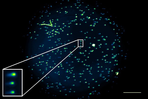
Optical mesoscopy image of single cell gel electro-phoresis assays. Prepared by Dr. Marie Boyd (Strathclyde Institute of Pharmacy and Biomedical Sciences, University of Strathclyde, UK)
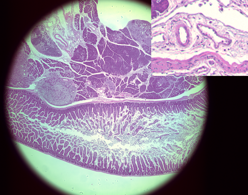
Mesoscope image of small intestine and liver tissue. The technology allows the visualisation of a single erythrocyte of less than 6 μm diameter within the 6mm FOV.
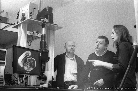
Dr John Dempster, Dr Brad Amos and Prof. Gail McConnell with the mesoscope